In emergency medicine, physicians use a broad array of diagnostic testing available to help patients who come to the ER with almost any problem. Technology has improved to upgrade various types of testing, especially imaging.
CT (Computed Tomography) or CAT (Computed Axial Tomography) is a quick imaging procedure that can significantly cut the time to diagnosis. Much of what you can see on a CT scan wasn’t visible outside surgery in days past.
Here is an overview of what a CT scan does, how it works, and when emergency departments deploy it.
Defining the CT Scan
A CT scan is a specialized X-ray procedure. Doctors use it to visualize bones, soft tissue, and blood vessels. The scan produces high-resolution images in cross-section, which the computer formats into a three-dimensional or multiple flat plane image.
CT scans help find injuries and diseases that aren’t readily visible to the naked eye and can’t be found through blood tests or traditional X-rays. They are accurate tools for quick diagnosis of issues in the pelvis, chest, abdomen, and other parts of the body.
The test is an X-ray beam that circles one part of the body, taking images from multiple angles. A computer combines the images to make cross-sectional pictures of the internal organs, bones, soft tissues, or blood vessels. The system allows doctors to view the images slice by slice if needed.
In some instances, the CT scan requires contrast media, which the patient swallows or has injected into the body. Contrast media helps the image more clearly define structures such as blood vessels, the urinary tract, the gallbladder, the liver, and the digestive tract.
What Is It Like to Have a CT Scan?
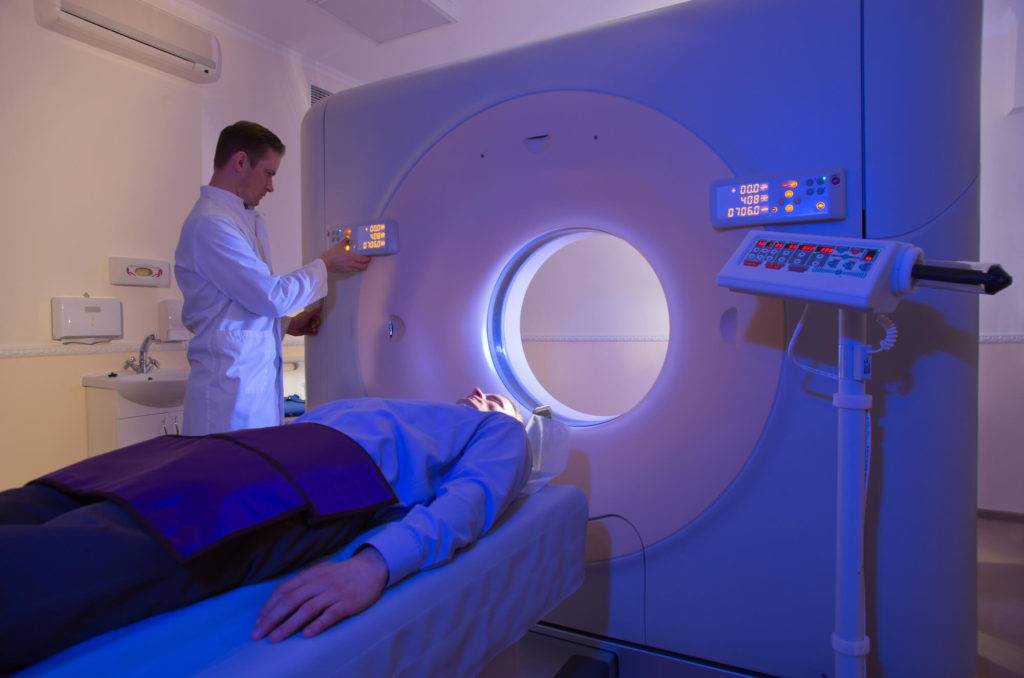
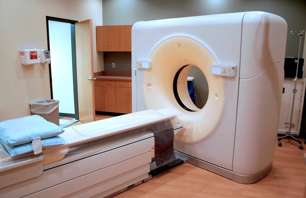
For the patient, the experience is simple. You lie on a platform that moves your body through a tube or donut-shaped structure. It’s painless and non-invasive (unless you require injected contrast, then just a little pinch).
Before the scan, you remove everything that could obscure an X-ray image, including jewelry, dentures, zippers, and other dense objects. During the scan, you must remain motionless so that the CT scan can capture clear images.
The entire procedure can take up to 30 minutes, depending on how extensive the image is. Some CT scans take only a few minutes. Once the scan is complete, a radiologist reads it and consults with the emergency room physician to help diagnose your problem.
Are There Risks to Having a CT Scan?
The risk due to X-ray exposure is not zero, but it is extremely low, no more than traditional X-rays. Some people are allergic to the contrast material used in some CT scans. They develop a rash or begin to itch but rarely experience anaphylaxis (an extreme reaction).
If you are pregnant, tell the doctor and X-ray technician before the procedure, so they can discuss whether the need for a CT scan causes problems for the fetus.
People with diabetes who take Metformin may be asked to stop briefly before and after the procedure in non-emergency situations, but for emergency purposes, you don’t need to worry about it.
Typical Emergency Use of the CT Scan
You might be surprised to learn that doctors take CT scans in the case of potential broken bones. While traditional X-rays are helpful in these cases, a CT scan provides more detail than conventional X-rays.
CT scans are used for detecting internal injuries and bleeding. The test shows both the severity and the extent of the injuries quickly. They are also used for stroke detection and treatment. It shows if there are bleeding or clots in the brain in time for the healthcare provider to provide fast treatment when time is of the essence.
Other diseases and injuries found using CT scans include:
- Appendicitis
- Kidney and bladder stones
- Injuries to the abdominal organs, such as the liver, spleen, and kidneys
Emergency medicine also uses CT scans to diagnose a pulmonary embolism, which is a blood clot in the lung. They can diagnose heart disease if the image shows blood clots that can cause a heart attack or excess fluid surrounding the heart and other internal organs.
A CT scan quickly visualizes bowel obstructions, tumors, and other abdominal disease processes. It can also be used to view infections of the soft tissues of the face and neck or lung infections and collections of fluid or air in the lungs.
In short, CT scans are used to help diagnose a wide variety of illnesses and injuries.
CT Scan vs. MRI
A CT scan uses X-rays to create an image, while an MRI uses magnets and radio waves. An MRI is better suited to imaging brain tissue, tendons, and ligaments than a CT scan and is also better for viewing the spinal cord.
An MRI is less valuable than a CT scan for diagnosing cancer, abnormal chest X-rays, pneumonia, and bleeding in the brain, especially after an injury. It shows organ tears and injuries more quickly and is more suitable for trauma diagnosis and care.
In Summary
CT scans have become the workhorse of emergency medicine when the speed of diagnosis requires quick yet precise testing. Computers have enabled CT scans, which are made from repeated X-rays taken from different angles, to provide slice-by-slice images or a 3D image of a bone or organ.
CT scans don’t hurt, though you may be asked to drink contract fluid or have it injected into a vein. Otherwise, you just relax and lie still. The emergency staff uses your CT scan for more definitive diagnoses than a medical history and visual check can provide.
CT Scans at Family First ER
The CT scan is part of the arsenal we use against injury and illness. We can find bleeding, broken bones, heart issues, and many other problems quickly so we can provide treatment as rapidly as possible.
When you or a loved one is experiencing a medical emergency, you want to be treated at a facility like Family First ER that has a complete line of future-forward diagnostic technology that help doctors quickly and accurately identify damage for precision treatment.